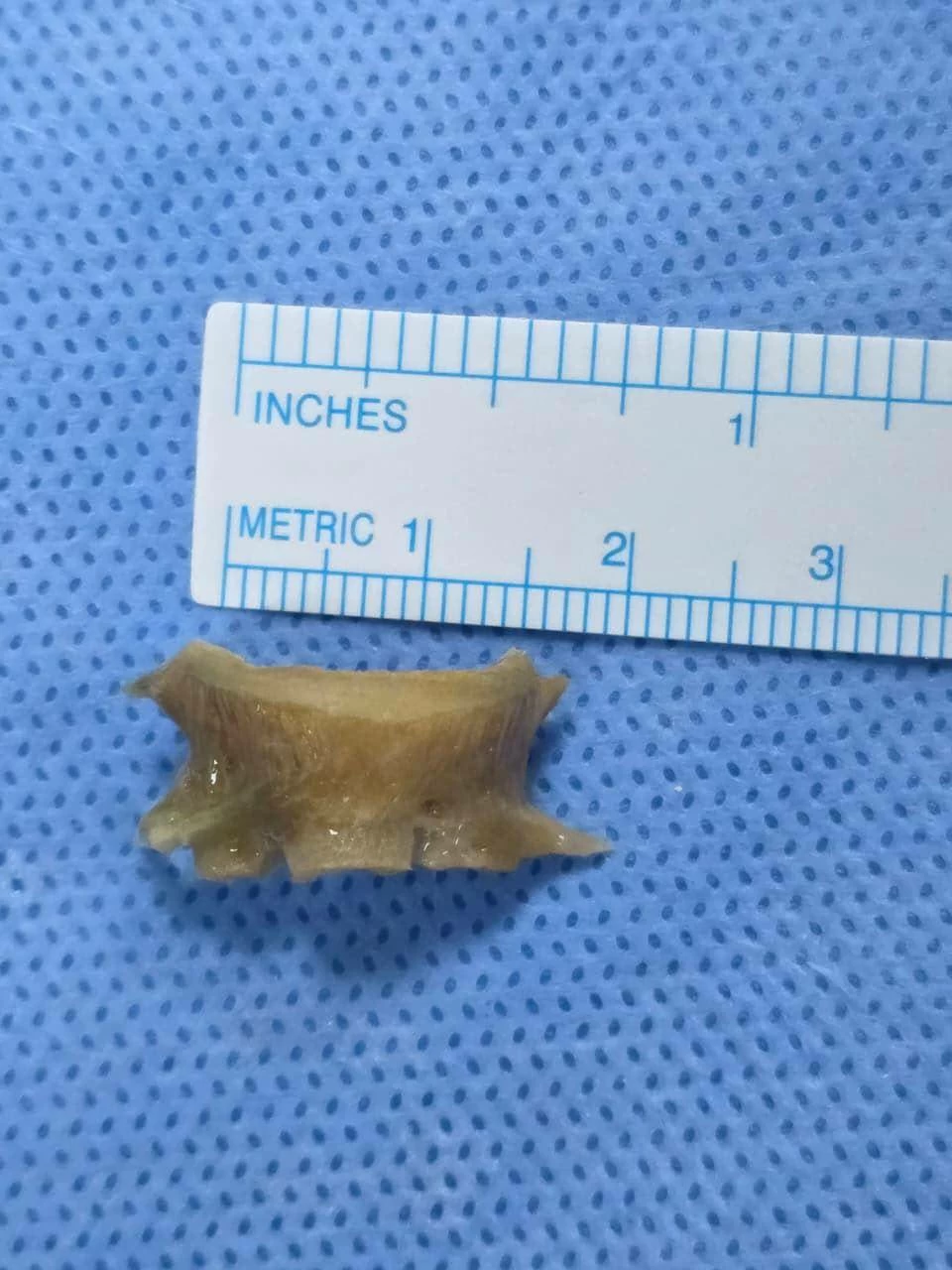
Dubai: Doctors at Aster Hospital Mankhool successfully removed a nearly 2-cm fish bone that had remained lodged in a patient’s lower airway for almost two months, after the 60-year-old patient experienced persistent cough, chest congestion and increasing difficulty in breathing following the accidental aspiration.
Fish bone foreign bodies are among the most common food-associated foreign bodies seen in adults, with a higher incidence reported in Asian populations. However, in this case, the fish bone had been accidentally aspirated into the lower airway and remained lodged in the vertical airway leading to the right lung for almost two months, resulting in significant local inflammation and granulation tissue, requiring a carefully planned multidisciplinary intervention.
Unnikrishnan Sivaraman, a 60-year-old Indian expatriate living in Sharjah, accidentally aspirated a milkfish bone while eating on July 1, 2026. Following the incident, he experienced an uneasy sensation, significant nausea and blood-stained vomiting and visited the emergency department at Aster Hospital Al Qusais (AHQ).
As his symptoms continued, including persistent cough, chest congestion and increasing difficulty in breathing, he made further medical consultations. A subsequent CT scan at another hospital identified the fish bone impacted in the lower airway. He was referred to AHQ for further management and was subsequently referred to Aster Hospital Mankhool as the case required specialised intervention.
The patient was referred to Aster Hospital Mankhool on August 26, 2026. Before the procedure, the Aster Hospital Mankhool team reviewed the CT images and conducted a further assessment, confirming that the fish bone remained lodged in the right main bronchus. Its prolonged presence had resulted in significant local inflammation and the formation of granulation tissue around the foreign body.
Given the size, sharp edges and location of the fish bone, its removal required careful planning. There was a potential risk of injury to the bronchial wall and surrounding structures, bleeding, sudden airway obstruction and difficulty maintaining adequate oxygenation during manipulation and extraction.
A multidisciplinary team from Pulmonology, ENT and Anesthesia planned the procedure, taking into consideration the location, sharp edges and prolonged presence of the foreign body.
(Left to right): Dr. Siraj Madayi Meethel, Specialist Anaesthesiologist; Dr. Jolsana Augustine, Specialist Pulmonologist; Mr. Unnikrishnan Sivaraman, patient; Dr. Bharath Neranki Satish Rao, Specialist ENT; and Dr. Abhishek Jayaprakash Narayan, HOD and Specialist Anaesthesiologist.Dr. Jolsana Augustine, Specialist Pulmonologist, performed bronchoscopy with anesthesia support from Dr. Abhishek Jayaprakash Narayan and Dr Siraj Madayi Meethel, carefully mobilised the fish bone from the right main bronchus towards the upper airway. Dr. Bharath Neranki Satish Rao, Specialist ENT, then performed direct laryngoscopy and used specialised forceps to remove the foreign body while protecting the vocal cords. A check bronchoscopy was subsequently performed to assess the airway and clear secretions.
Dr. Jolsana Augustine, Specialist Pulmonologist, Aster Hospital Mankhool, said, “When a foreign body remains in the airway for a prolonged period, inflammation and granulation tissue can make removal more challenging. In this case, the size, sharp edges and location of the fish bone required careful planning and controlled manipulation. Close coordination between the Pulmonology, ENT and Anesthesia teams was essential for its safe removal while minimising the risk of airway injury or bleeding.”
Dr. Bharath Neranki Satish Rao, Specialist ENT, Aster Hospital Mankhool, said, “The sharp edges of the fish bone and its proximity to the vocal cords made the final stage of extraction particularly delicate. Our focus was to remove the foreign body safely while protecting the vocal cord structures. The successful outcome was made possible by careful coordination between the Pulmonology, ENT and Anesthesia teams throughout the procedure.”
The procedure was completed successfully on 11th September 2026, and the patient was discharged home in a stable condition within a few hours. At follow-up, he reported significant improvement and was doing well, with no further medical treatment required.
Expressing his gratitude to the medical team, Sivaraman said, “I am very thankful to Dr. Jolsana, Dr. Bharath and the entire team at Aster Hospital Mankhool for the care and support I received. Dr. Jolsana explained everything clearly and made me feel comfortable throughout the treatment. I am very happy with my recovery and the significant improvement in my breathing and overall condition.”
The case highlights the importance of accurate diagnosis, careful pre-procedure planning and multidisciplinary teamwork in managing complex airway foreign bodies, particularly when the foreign body has remained lodged for an extended period.